
Diffraction
Structural investigations of SURMOFs
Different set-ups resp. diffractometer are available: Out-of-plane (coplanar) in θ-θ geometry: determination of lattice plane spacing parallel to the substrate, e.g., (00l)-planes with l=1,2,3.
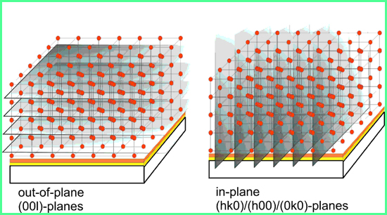
Figure 1: The orientation of the planes detectable for the different set-ups
In-plane (non-coplanar) geometry: determination of lattice plane spacing perpendicular to the substrate, e.g., (hk0)-planes with h,k,=1,2,3.
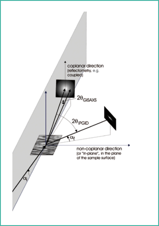
Figure 2: The geometry of the in-plane configuration (Picture from LAB Report XRD 68, Bruker AXS).
Examples for the OP and IP diffractograms of a HKUST-1 SURMOF are depicted in figure 3 and 4.
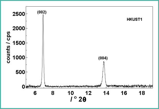
Figure 3: Out-of-plane diffractogram of an HKUST-1 SURMOF. This is also the first quality check in the SURMOF production.
Dies ist auch der erste Qualitätscheck in der Herstellung der SURMOF. (Abb3.jpg)
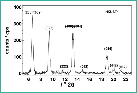
Figure 4: In-plane diffractogram of HKUST-1. Clearly more peaks are visible. This additional information is essential for the structure determination e.g., with Rietveld methods
Diese zusätzliche Information ist sehr wichtig füreine Strukturbestimmung , z.B. mittels Rietveldmethoden.
In-Situ investigations of SUFMOFs
Monitoring of the intensity chance of certain Braggpeaks reveals the kinetics of the loading resp. incorporation of guest-molecules into the SURMOF pore structure.
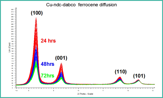
Figure 5: Intensity change, especially of the first order Braggpeaks with increasing incorporation of guest molecules into the SURMOF lattice.
Heating the SURMOF in special heating-stages and simultaneously recording the diffractograms allows to determine physical properties of the SUR-MOFs. The example presents heating of HKUST-1 from RT to 150°C. Data evaluation yields a negative thermal expansion factor, i.e. the dimension of the units cell shrinks while heating.
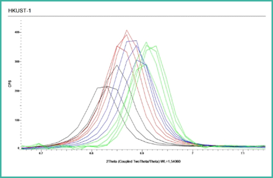
Figure 6: Shift of the Braggpeak position to higher angles with increasing temperature.
X-Ray Reflection (XRR)
Moreover X-ray reflection can be applied in order to determine the layer-thickness and layer composition of the sunstrates.
This video: ![]() from Bruker-AXS presents a very comprehensice introducttion into grazing incidence diffraction (GID)
from Bruker-AXS presents a very comprehensice introducttion into grazing incidence diffraction (GID)
https://my.bruker.com/acton/ct/2655/s-0ea0-2004/Bct/l-16db2/l-16db2:1664d/ct19_2/1?
sid=TV2%3AGn21skbii