


Platelets are anuclear 3 – 4 µm cell fragments circulating in blood. They play a crucial role in haemostasis, the process of blood coagulation, as shown in the figure below.
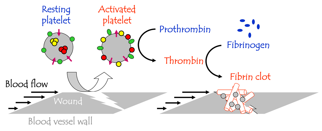
| Platelet activation at the wound site. Platelets are shown as large grey spheres on the left hand side. They circulate in the blood in an inactive (quiescent) form. They undergo activation at the wound site (e.g., tear in the blood wall). Activated platelets catalyze clot formation (right hand side) by converting on their surface prothrombin into thrombin, which then converts fibrinogen into fibrin, that forms the clot. The clot contains platelets (small grey spheres in the right hand side image) as well as other blood cells (not shown here but see below). |
Other platelet functions and selectivity
Recently, multitude of new platelet functions have been discovered.[Leslie M. 2010, Science 328, 562] They play an important role in wound healing, inflammation, innate and adaptive immune response, and participate in rejection or integration of artificial implants.
When activated, platelets also secrete over 200 active substances – growth factors and cytokines – with various, sometimes contradictory functions.[4] Understanding how the secretion of these factors is regulated would allow the regenerative potential of the platelets to be harnessed.
In my group, we are studying platelet activation
- by biomaterials (in the context of biocompatibility)
- by natural agonists (in the context of physiological coagulation and wound healing as well as pathological thrombosis)
- In whole blood, platelet-rich plasma, and purified platelets
Our goals are
- To design new assays for studying platelet activation kinetics
- To develop model systems for identifying different functional states of platelets
- To advance our knowledge of platelet signaling pathways so that we can tune platelet responses to improve implant integration and design better antiplatelet therapies.
Example of selective platelet activation by surfaces
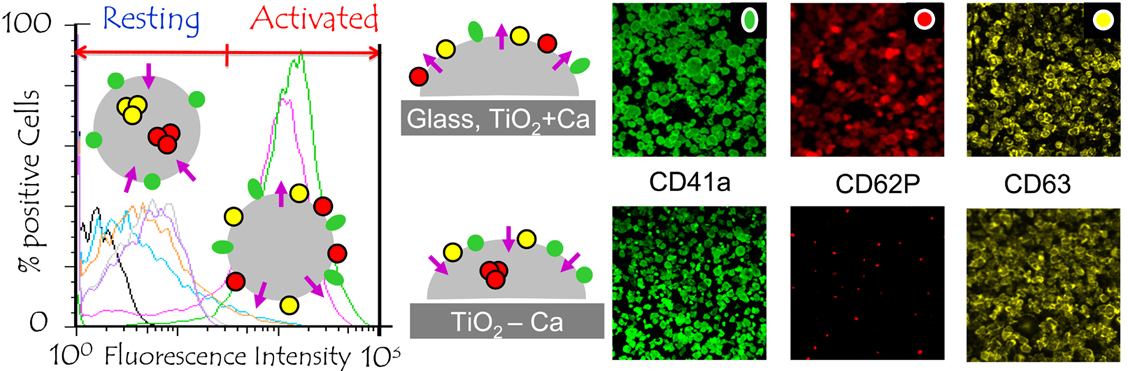
| Platelet activation profiles on the surface of a popular biomaterial, TiO2, depend on whether Ca2+ is present or not. overlaying the flow cytometry data (left). Fluorescent images of adherent platelets are shown on the right. In solution, activated platelets can be distinguished from the resting platelets by flow cytometry (left). Platelets in solution are shown as large grey circles. For platelets adhering at surfaces, the same can be done by fluorecence microscopy (right). Adherent platelets are shown as semi-circles (middle). Activated platelets are distinguished from the resting platelets by labeling them with antibodies against specific markers: CD63, dense granule marker (small yellow circles). CD62P, alpha granule marker, small red circles. Phosphatidyl serine, purple arrowheads. Activate form of the integrin complex GPIIb-IIIa, green ellipse. Its inactive form, CD41a, expressed on the resting platelets, is shown as a green circle. Resting platelets do not express PS, CD62P, or CD63. Activated platelets in solution, platelets adsorbed on glass, or on TiO2 in the presence of Ca2+, express all these markers. Platelets adsorbed on TiO2 in the absence of Ca2+ do not express CD62P. Selectivity of platelet activation is a topic of considerable current interest due to the possible applications in implant integration, thrombosis and bleeding therapies, and drug delivery protocols. |
References:
Gupta, S. / Reviakine, I. (2012): „Platelet Activation Profiles on TiO2: Effect of Ca2+ Binding to the Surface“. In: Biointerphases (2012), 7, 28 – 40.
Gupta, S. (2012): „Selective Activation of Platelets by Surfaces and Soluble Agonists“. In: Ph.D. thesis, The University of the Basque Country, Bilbao, Spain, 2014.
Further information can be found on my website, www.ireviakine.net
