
ESEM
All microscopic measurements are carried out in a FEI Philips XL 30 Field Emission Gun Environmental Scanning Electron Microscope (FEG-ESEM). As one of the basic characterization methods at the institute, electron microscopy is used for the investigation of samples from numerous research fields, e.g. nanomineralogy (layered minerals, mesoporous materials), polymer science, investigation of cement phases, or metal organic frameworks. (Fig. 1)
In most of the cases a complex sample preparation is not necessary. Nevertheless, to avoid charging, insulating materials have to be coated with a thin layer of conductive carbon or with a Gold/Palladium-film. Then the specimen can be imaged under high vacuum conditions (10-5 Pa) using acceleration voltages between 5 and 20 kV.
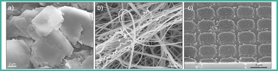
Figure 1. Examples of investigated materials: a) layered minerals and mesoporous materials, b) polymers and nanocomposites, c) metal organic framework structures (MOF).
Furthermore it is possible to run the ESEM under various pressures between 100 and 1300 Pa. Due to its special design sensitive samples like organic and biological substances can be imaged without complex drying procedures and without a conductive coating, which minimizes beam damage. (Fig. 2)
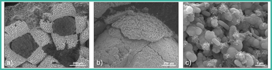
Figure 2. Images of vacuum sensitive biological samples, a) spores and fungi, b) biofilm, c) bacteria.
A tensile stage (M-TEST300, Deben Co.) can be used to run in-situ tensile test (stress/strain experiments). During these experiments the ductile behavior of the materials can be investigated, as well as morphological changes (cracks). (Fig. 3)
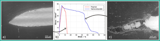
Figure 3. Tensile tests on polymer and nanocomposite samples, a) brittle failure of the raw polymer (red curve), b) stress-strain diagram, c) ductile failure of LDH-nanocomposite with strongly increased tensile strength (blue curve).
Elemental analyses and elemental mapping are performed using the energy dispersive X-ray fluorescence spectroscopic unit (EDX) from EDAX with a liquid nitrogen cooled Sapphire Si(Li) detector. For the spectra collection the microscope was operated a 100 µm aperture was used. With this setup point spectra or linescans can be recorded, as well as elemental mappings over larger sample areas. (Fig. 4)
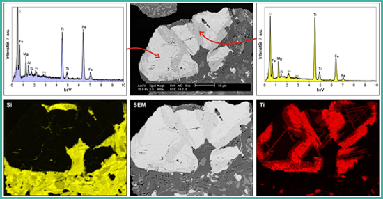
Figure 4. . Energy dispersive X-ray spectrometry (EDX). Top) single spectra recorded in the light and dark areas of the grain; bottom) elemental mapping of the entire area.
Equipment
Device Designation: Philips XL30 ESEM-FEG (FEI)
Classification: Environmental Scanning Electron Microscope with Field Emission Gun (ESEM-FEG)
Description/Features:
- Detectors: Everhart-Thornley secondary electron detector (SE), backscattered electron detector (BSE), several gaseous secondary electron detectors (GSE’s), from large-field detector to 0.5-mm-aperture detector
- EDAX liquid nitrogen cooled energy-dispersive X-ray spectrometry (EDX)
- Peltier heating/cooling stage
- Bal-Tec MED020 cryopreparation, incl. shuttle transfer system and cryo-stage.
- Coating of Au/Pd 80/20 (Bal-Tec MED020), and Carbon (Cressington 108carbon/A)
- Tensile Stage MTEST300 (Deben/Gatan)
Keywords: SEM, ESEM, variable pressure microscopy, sensitive samples.